Review Article
Digging Back in Evolution: Danger in Drosophila
Alexis Dziedziech, Dilan Khalili and Ulrich Theopold*
Corresponding Author: Ulrich Theopold, Department of Molecular Biosciences, The Wenner-Gren Institute, Svante Arrhenius väg 20c, S-10691 Stockholm, Sweden
Received: July 19 2018; Revised: January 10, 2020; Accepted: July 23, 2018 Available Online: October 20, 2018
Citation: Dziedziech A, Khalili D & Theopold U. (2020) Digging Back in Evolution: Danger in Drosophila. J Cell Signal Damage Assoc Mol Patterns, 1(1): 1-8.
Copyrights: ©2020 Dziedziech A, Khalili D & Theopold U. This is an open-access article distributed under the terms of the Creative Commons Attribution License, which permits unrestricted use, distribution, and reproduction in any medium, provided the original author and source are credited.
Share :
  • 1008

    Views & Citations
  • 8

    Likes & Shares

Insects, including the fruit fly, Drosophila melanogaster are used to study a wide array of processes, many of which are known or are expected to be regulated by damage-associated molecular patterns (DAMPs). These include regenerative processes after wounding, replacement of cells by cell competition, induction of immunity and inflammation, responses against tumorous cells and neurodegeneration. Most, if not all of these processes have beneficial outcomes on organismal health but may also lead to pathologies, which often resemble those observed in humans. Drosophila offers unique opportunities to analyze and manipulate genes and pathways related to these immune consequences with high temporal and local resolution. Ultimately, such detailed analyses in the Drosophila model will aid in our understanding of the roles DAMPs play at the bifurcation between physiological and pathological outcomes in other animal species, including humans.


Keywords: Coagulation, Danger signals, DAMPs, Hemocytes, Inflammation, Innate immunity, Insect immunity, Regeneration, Tumors, Wound healing


Abbreviations: DAMPs: Damage Associated Molecular Patterns; MAMPs: Microbe-Associated Molecular Patterns; Egr: Eiger; TLRs: Toll-Like Receptors; ITAMs: Immunoreceptor Tyrosine-Based Activation Motif; AMPs: Antimicrobial Peptides; Drs: Drosomycin; PPO: Prophenoloxidase; Spz: Spätzle; PRRs: Pattern Recognition Receptors; ROS: Reactive Oxygen Species; Crt: Calreticulin; MMP: Metalloproteinase-1; EPN: Entomopathogenic Nematodes; Psh: Persephone; ECM: Extracellular Matrix; ER: Endoplasmic Reticulum

INTRODUCTION

 
Insects serve as models which not only expand our understanding of innate immunity, but also shed light on mammalian immunology. While differences between vertebrates and spineless creatures have arisen evolutionarily due to distance and varied ecological niches, a vast number of similarities can be found between these two groups. Furthermore, insects offer methods and tools that constitute a complementary investigative approach for innate immunologists. Here, we provide an updated summary on danger and damage signals in insect immunity as previously reviewed [1] and focus primarily on research performed on the fruit fly, Drosophila melanogaster but occasionally refer to other models. One notable example for the success of fruit fly immunology is the identification of the immunological role of Toll signaling, which paved the way for discovering analogous pathways in vertebrates [2]. Conversely, when it comes to Toll signaling, a number of damage associated molecular patterns (DAMPs) have been identified to be recognized by members of vertebrates’ Toll-like receptors (TLRs) while similar roles for fly Toll signaling are only just emerging (Figure 1). However, through the use of the expansive Drosophila tool box, immunologists have been able to further elucidate the complexity of the innate immune response, which exists beyond recognition between self and non-self through the ability to discriminate between normal host factors and endogenous danger signals. Danger signals can be tissue specific, recognized by a downstream DAMP factor based on: a) microbial threat; b) traumatic injury; or c) sterile inflammation or tumor development. Several mammalian DAMPs have been identified ranging from single nucleotides to proteins. Similarly, in Drosophila melanogaster there is accumulating evidence for conserved DAMPs. Conserved DAMP factors that have been demonstrated to play a role in both sterile and septic injuries are Actin, ATP, Calreticulin, Egr (TNF-α) and Reactive Oxygen Species (ROS). Interestingly, other non-homologous proteins have also been recognized as DAMPs in the fruit fly, such as Spätzle (Spz), which has only been observed to be active in septic wounding situations.

DROSOPHILA IMMUNITY - A QUICK GUIDE

 
The fruit fly, Drosophila melanogaster—like all invertebrates—lacks the prototypical adaptive immune response yet is nevertheless capable of mounting highly effective innate immune responses which include humoral factors released by the fat body (the insect liver equivalent) into the hemolymph (the insect blood equivalent) and specialized immune cells called hemocytes which are most akin to cells of the mammalian myeloid lineage (hemocytes) [3]. Many insect immune reactions rely on a combination of humoral and cellular activities. Invertebrate humoral factors are comprised of variable cocktails full of antimicrobial peptides (AMPs, of which Drosophila contain 8 classes), proteins with similarity to complement components, members of a proteolytic cascade that ultimately activate the zymogen prophenoloxidase (PPO), the clotting system which seals wounds and prevents dissemination of microbial intruders [4] and finally, a wide array of proteins with unknown function. Two pathways (Toll and imd) are classically activated by microbial elicitors and are required for the induction and release of many immune effectors. Both pathways are traditionally thought to be activated by bacterial elicitors (Lys-type peptidoglycan and DAP-type peptidoglycan, respectively) and lead ultimately to the activation of NFkB-like transcription factors into the nucleus. Toll activation requires extracellular activation of the Spätzle (Spz) ligand either downstream of pattern recognition receptors (PRRs) with specificity for bacterial microbe associated molecular patterns (MAMPs) such as peptidoglycan and fungal glycans or downstream of DAMPs. Two stress-related pathways (JAK/STAT and JNK) contribute to immunity in different ways depending on the tissue/cellular and developmental state. While hemocytes (blood cells) in Drosophila can contribute to the downstream effects of the aforementioned humoral responses, they also contain their own specialized cellular immune functions. Hemocytes activate upon different wounding scenarios and include plasmatocytes which embody both macrophage and granulocyte properties, crystal cells which harbor PPO which is involved in melanization at the wound site and lamellocytes, a third class of larger flat cells which encapsulate larger intruders and thus primarily differentiate upon the detected presence of these invaders [3]. With all the complexity of the innate immune system to still be uncovered, being able to focus on any aspect of immune regulation by damage/danger signals in the fly inherently allows us to parse out the key functions which give rise to a better understanding of the innate branch of immunity. From the fly, we have learned that the innate immune system does not purely activate in response to exogenous danger signals like those elicited from pathogens, but also in a way that can mirror exogenous activation but by endogenous factors. This aspect of immunity has been dubbed “sterile inflammation” [5]. In this review, we will discuss examples in which both microbe-induced and endogenously released DAMPs collectively contribute to and inform immunity in the fly (Figure 2) and vertebrates, respectively.

WOUND SEALING AND HEALING: LIVE IMAGING AND BEYOND

 
One of the most obvious places one could expect to see DAMPs is at a wound site. Wounds can be studied via mechanical damage with lasers or surgical needles, injections with microbes, infections with parasites or via sterile wounds, such as pinching and other means of mechanical stimulation that have been established and exclude microbial factors [6-8]. Sterile wounds avoid clot formation but often recapitulate the events transpiring after an external wound occurs, like in the case of needles and lasers. In Drosophila, reactions at the wound site can be easily followed in vivo due to the translucent nature of most of its developmental stages, which allows for live imaging using fluorescent markers and the vast array of existing tools for fly genetics [6]. Live imaging has played a major role in elucidating the sequence of events at epithelial wounds but also at early embryonic syncytial stages, which serve as models for cellular wounds [9,10]. In most of these stages, wounds are quickly sealed by a hemolymph clot, which often forms within seconds after the tissue is damaged. In this brief window, rearrangements of the cells that surround the  wound  occurs  and  variably  involves  the  formation of  In Drosophila, the secretory DAMPs that are best characterized are Actin, ATP, Calreticulin (CRT), ROS, Eiger (Egr) and Spätzle (Spz). During tumor growth or cell injury, the cell becomes inflamed and as a result, releases DAMPs such as actin which may be exposed at the injury site (red circles) or released into circulation. When actin is in circulation, it may act as a DAMP, to induce a DAR (DAMP Associated Response), as do ATP, CRT, ROS, Egr and Spz. These DARs may include the activation of plasmatocytes and/or crystal cells and/or the JAK/STAT pathway in the fat body which can result in the production and release of cytokines. Similarly, accumulation of Actin, ATP, CRT, ROS, Egr and Spz in hemolymph circulation may result in the activation of hemocytes to promote wound healing
filopodia, lamellipodia and transcellular actin-cables, all of which aid in closing the epithelial gap. While there is still much to unveil in respect to how wounds recruit cells and heal, increased temporal and local resolution combined with computational modeling of the inflammatory response to tissue damage has provided further insight into the molecular events at the wound site. Weavers et al. [11] found that the kinetics of diffusion of the wound signal appears incompatible with a compound of low molecular mass due to the fact that the rate of diffusion is theoretically too slow for small molecules to be the primary chemoattractant. This may disqualify classical DAMPs such as ATP and H2O2, both of which might instead indirectly contribute to a pro-inflammatory environment. Modeling and experimental confirmation of multiple wounds further identified a temporal window after wounding during which hemocytes are insensitive to the wound signal implying there may be a ‘switch’ like that which is known for G-protein-coupled chemokine receptors. In addition, hemocytes displayed heterogeneity in responses that may ultimately aid in understanding the difference between acute-transient and chronic-inflammatory wound healing [11]. Though the kinetics are being pieced together, it is still unknown how activation of the clot initiates (i.e. what the tissue factor equivalent is), but it is known that reactive oxygen species (ROS) play a major role during wound healing. ROS can injure pathogens, stimulate hemocytes and induce a systemic response [12]. H2O2 acts as an evolutionary conserved damage signal in both zebrafish and Drosophila acting on neutrophils and hemocytes, respectively by creating chemokine gradients which attract immune cells [13-16]. The signal transduction cascade that is activated downstream of H2O2 is conserved from fruit flies to vertebrates and has likely endured genetically based on its role in healing insect wounds, which is to distinguish self from non-self [15]. Live imaging in combination with laser wounding has identified a conserved cascade (SFK-Draper-Shark) downstream of H2O2, which comprises Draper, an apoptotic clearance receptor containing an ITAM (immunoreceptor tyrosine-based activation motif) domain, which plays a crucial role in recruiting cells to the wound site. Draper is activated by a Src family kinase (SFK; in Drosophila Src42A) and activates a downstream kinase of the Syk family (Drosophila Shark) required for hemocyte migration to the wound [15].

In an infection context, hemolymph clots have been demonstrated to prevent entry of parasites that target epithelial surfaces using mechanical tools such as entomopathogenic nematodes (EPNs), which use their mouth part to gain entry to the hemocoel [4]. EPN infection leads to a massive induction of immune-related genes although some immune genes appear to depend on the clot or clot components for their induction rather than on microbial or parasite-specific elicitors [17] and thus are more akin to microbe-independent responses like those observed during sterile inflammation. In microarray data of Drosophila infected with the EPN Heterorhabditis bacteriophora, it was found that several hundred genes are specifically induced in EPN infection in comparison to other types of infections, like parasitic wasp infection, and that there are several candidate damage-induced molecules such as thioester containing protein-1, Eiger, Spätzle-processing enzyme and potentially others that are yet to be characterized [17,18]. Interestingly, the Toll reporter Drosomycin is highest on that list providing evidence for alternative ways of inducing AMPs independent of the Toll pathway. Similarly, Hauling et al. [19], through RNA sequencing of the fat body gained further insights into the endogenous response against danger signals produced from tumor-expressing salivary glands [19]. Beyond the current molecular scope, we have successfully been implementing the use of sequencing data as a map for finding new danger signals and DAMPs.

 
ACTIN AND DANGER

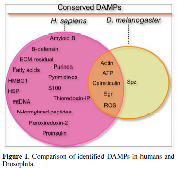
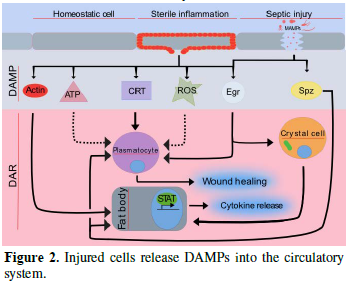
Released DAMPs enable broad trans-tissue communication. In external wounding situations, local DAMPs at wound sites establish local communication between the wound and the immune system to ensure that a proper wound healing response is taking place. In injured organs, actin can be exposed on the surface of the wound site [20]. Similar findings have revealed that actin is also found in the blood, suggesting that it may be released from injured tissue [21]. However, the mechanism of DAMP-actin-induced inflammation is not fully understood. Another role actin may have is to serve as a signal molecule [22]. For instance, Ahrens et al. [23] found that actin is a danger signal that is conserved from yeast to humans. F-actin acts as a ligand and is recognized by a DAMP receptor for dead cells called DNGR-1 (also known as CLEC9A) in both vertebrates and Saccharomyces cerevisiae [23]. In mosquitoes, actin has been found to promote phagocytosis of bacteria in cooperation with the small MD2-like protein and to act as a Plasmodium falciparum antagonist [22]. Recently, Srinivasan et al. [24] identified actin as a conserved DAMP in the fruit fly. Following injection of actin into Drosophila larvae, they observed induced sterile inflammation in the fat body. They identified that exogenous actin acts as a conserved signal that is released from damaged cells which leads to a selective JAK/STAT response [24]. While the actual actin receptor remains elusive (there is no fly homologue for the mammalian actin receptor DNGR-1 and Draper appears dispensable), the actin signal appears to feed into the SFK-Draper-Shark pathway further strengthening its evolutionary conservation as a cell injury detector which precedes the evolution of adaptive immunity.

NON-CANONICAL TOLL SIGNALING: A VERSATILE TOOL [25]

 
An oft-used tool to determine induction via Toll is the expression of the AMP, Drosomycin (Drs), which can be checked by using qPCR or Drs reporter lines. In this way, Drs activation has been observed even in the absence of microbial elicitors and in some cases involves Toll and its ligand, Spätzle, but in other cases appears to be activated in a Toll-independent manner [8]. In an apoptosis-defective setting, induction of immunity requires the presence of the protease detector, Persephone (Psh), the extracellular Toll sensor, Spätzle and the translocation of the NFkB-like transcription factor, Dorsal [26]. When using a partially purified larval extract, Kanoh et al. [27] found strong Drs activation via the Toll receptor [27] however, RNAi screening identified an additional non-canonical transcription factor, Jarid2 (a Jominji-like transcription factor), which was required for full Drs induction. In addition, Spz4, which is related to canonical Spz may also have contributed to Toll activation. While the DAMP in the extract remained elusive in this study, another study found that in the absence of immune attack, Spz can be activated in Senju mutants. Senju encodes a UPD-galactose transporter and in its absence the penultimate carbohydrate will be exposed and may act as a potential DAMP [28]. Toll can therefore be activated and negatively regulated in a broader range of scenarios than traditionally described [25]. Non-classical Toll signaling also includes activation of a well-studied phenomenon in Drosophila namely cell-competition via additional members of the Drosophila Toll-family. During cell-competition, unfit cells (loser cells) are eliminated by their normal neighbors (winner cells) to prevent them from being a part of the mature tissue. An unfit status can be induced experimentally when cells carry a minute mutation, which affects ribosomal proteins and cell growth. On the other hand, cells may achieve a competitive advantage over normal neighbors (super competitors cells), for example by increased expression of the myc oncogene. Both scenarios were studied and shown to induce apoptosis in the loser cells, in both cases relying on Spz and on different members of the nine Drosophila Toll-related receptors (TRRs) [29]. TRRs 3 and 4 conferred a loser status to minute cells and TRRs 2, 3, 8 and 9 conferred a loser status to wild type cells adjacent to myc-expressing cells. The two environments required different modules of both Toll and imd signaling and also different NFkB members (Dorsal/DIF and Rel, respectively) but both still led to apoptotic death of loser cells. Thus, in this scenario innate immune modules are essential in maintaining tissue integrity through the elimination of unfit cells.

PERSEPHONE: BAIT FOR EXOGENOUS PROTEASES AND ENDOGENOUS SIGNALS

 
To gain access to the host, microbial attackers have to breach the protective host cuticle often through the secretion of virulence factors such as proteases. These exogenous microbial proteases thus act as Danger signals which serve as proxies to indicate the presence of microbes via their (indispensable) invasive activity [30]. Interestingly, Toll signaling has been co-opted to detect such proteases. Instead of being classically activated through MAMPs from Gram-positive bacteria and entomopathogenic fungi, a protease (Persephone, Psh) upstream of Toll is sensitive to a wide range of microbial proteases which include Gram-negative bacteria. A bait region exposed on Psh contains a number of sequences that are sensitive to different classes of exogenous microbial proteases conferring wide-range protection. Thus, the bait region of Psh can respond to exogenous proteases regardless of whether or not the protease-producing microbes fall within the classical categorization of Toll being primarily activated by beta-glucans from fungal cell walls or lysine-type peptidoglycan from Gram-positive bacteria. Furthermore, Psh is indispensable for Toll activation in an in vivo fly model for defective apoptosis in which endogenous DAMP signals trip the bait region of Psh [26]. Although the activating mechanism is as of yet unknown, these combined findings place Psh at a central position upstream of Toll which leads to activation of immunity via detection of both exogenous and endogenous damage-inducing activities. 
 
TRAINED IMMUNITY: DOES IT DIFFER BETWEEN INSECTS AND MAMMALS?

In trained immunity in mammals, innate immune cells can be primed by primary infections or vaccination to perform more efficiently upon subsequent exposure to microbial attack. Similarly, insect immunity can be primed by previous exposure to antigens, a phenomenon that has been dubbed “immune memory” or “immune priming” [31]. While trained immunity in mammals confers broad range protection against unspecific microbes, at least some cases of insect immune priming appear to be quite specific [32]. Though the possibility that the mechanism exists in vertebrates and organisms with an adaptive immunity has not yet been ruled out. While exogenous signals have been found responsible for immune training/priming, similarly in mammals and insects, tissue damage appears to play a central educational role for insect hemocytes [33]. Like macrophages, hemocytes are multifunctional cells taking care of both internal damage and microbial attack by for example, phagocytosing bacteria or removing apoptotic cells. During the removal of apoptotic cells, Weavers et al. [33] showed that Drosophila embryonic development is essential for priming hemocytes both to efficiently perform wound healing and to fight infections. Priming is triggered by calcium flashes which activate JNK signaling and subsequent induction of the apoptotic regulator Draper, a key molecule in wound healing. Consequently, inhibiting apoptosis as well as interfering with JNK signaling affects the inflammatory potentials of hemocytes, which is somewhat expected but surprisingly, their immune competence is hindered for example, they lose the ability to phagocytose Escherichia coli [33]. Trained immunity has also been demonstrated in the case of viral infections in the fruit fly. Cellular damage releases viral dsRNA which is subsequently phagocytised by plasmatocytes and eventually packaged into endosomes to transfer antiviral RNAi to other hemocytes [34]. Thus the mechanism for priming hemocytes for a specific viral infection initially bears the hallmarks of damage-induced clearance but leads to specific protection. This varies widely from what has thus far been shown in the adaptive immune system in the mammalian model, but demonstrates that the innate immune system can play a similar role in immune priming.

CANCER AND DANGER: A MULTILEVEL AFFAIR

“Fail to heal” is one of the attributes in tumor developing tissues [35]. As a result, DAMPs may be released from tumor-derived wounds and act as pro- or anti-tumor factors [36]. Drosophila, despite a much shorter lifespan, has been used to successfully model the progression of human tumor growth and its consequences on neighboring cells and the immune system. Starting in the early 2000s, tumor growth in flies was induced either in a mosaic fashion using somatic recombination, in defined tissues by either overexpression or dominant-active forms of proto-oncogenes, or by decreasing the expression of tumor suppressors often in combination with the two other modes of tumor induction [37,38]. By combining this basic setup with potential regulators of tumor progression, modifier-screens identified several genes and pathways that were active either in the dysplastic cells themselves or in adjacent cells in the same tissue/organ. JNK signaling and the Drosophila equivalent of TNF, Eiger (Egr) were shown to modulate tumor growth. Both pathways had the potential for pro- or anti-tumor activity depending on tumor background and in which tissues they were expressed. In particular, when it came to studying the influence of adjacent cells on tumor progression, Drosophila became an excellent model due to its genetic toolkit. In addition, cell competition, which is akin to tumor surveillance within tissues, was a phenomenon already well-known to Drosophila geneticists. Using fly genetics to study the interaction between tumor cells and adjacent cells, studies showed that: a) in addition to supporting super competitors, cell competition is a homeostatic mechanism able to prevent tumor growth; b) apoptosis of cells (such as loser cells) may induce compensatory proliferation in neighboring cells, which paradoxically bears its own risk for aberrant growth due to chronic activation; and c) similar to many mammalian tumors the basement membrane is often degraded, facilitating invasive growth and metastasis [39,40].  
When inducing tumor growth in somatic tissues, it also became apparent that an immune response was transactivated in immune cells [19,41]. Both hemocytes and the fat body were found to activate a strong anti-tumor response with some similarities to their antimicrobial activity but also features specific to the tumor’s presence. It still remains necessary to pinpoint which factors are promoting the differential response between different cell types and wounds however, this evidence promotes the idea of DAMP-induced sterile-inflammation in Drosophila tumor models. Nevertheless, as a result, activated hemocytes are recruited towards tumors where they may have tumor-promoting or limiting activity. In particular, their pro-inflammatory potential, which is enhanced through the collaboration with the local response has been studied and involves ROS, JNK and TNF signaling [42], i.e., classical damage signals/pathways. Furthermore, retinoids may support tumor growth: flies that express a dominant-active form of the Ras oncogene and are defective in retinoid metabolism survive much better than flies expressing Ras alone [19]. If instead of expressing Ras, apoptosis is induced in the same tissues, retinoids play an anti-inflammatory protective role [43] and support transient wound healing. In other contexts, such as virus infections, retinoids may activate retinoid-induced genes as part of an antiviral response [44]. Another emerging DAMP factor, Calreticulin (CRT), a Ca2+ binding protein in the ER [45], has been found in the extracellular matrix (ECM) of rat predentin [46] and may contribute to phagocytosis in Drosophila. For instance, phagocytosis of ecdysone-induced apoptosis of S2 (macrophage-like) cells was reduced when CRT was blocked with α-CRT antibody. Similarly, in Drosophila embryos, a CRT knock-out reduced the number of phagocytosed cells [47]. Further studies are needed to understand whether Drosophila CRT can act as a DAMP in the ECM and induce sterile inflammation via hemocytes.

DANGER IN THE NERVOUS SYSTEM: NEUROINFLAMMATION

A number of approaches have been used to induce damage in the fly’s nervous system (NS) which include the expression of human disease-causing gene polymorphisms in the NS [48] as well as through causing mechanical damage [49]. Upon severing axons that lead to either wings or legs, transcriptome profiling of the ventral nerve cord revealed two main pathways involved in other wounding scenarios, mainly Draper/AP-1 and Toll, were upregulated. In addition, Stat92E/draper/JNK/AP-1 activity was necessary for metalloproteinase-1 (MMP1) to successfully clear away debris of severed axons and for regeneration [49]. While this study demonstrated activation of stress was required for innate glial immunity, another study used a fly model of Ataxia-Telangiectasia to demonstrate that the NFkB factor, Relish and the induction of select immune genes were key culprits for neurodegeneration [48] in a non-canonical manner (the imd pathway was dispensable). When different models of neurodegenerative scenarios are compared, an immune signature is often identified. It has been proposed that this immune signature may actually reveal an equally important neuroprotective function of the “immune genes” since both DAMPs and damage clearance play key roles during infections and tissue healing, a pleiotropy that is often misrepresented during gene annotation [50,51]. An example of this duality which exists within genes’ function is provided by a member of a prototypical PRR family (PGRP-LC, a peptidoglycan receptor in the Drosophila imd pathway), which is also required for synaptic plasticity in mice [52]. Similarly, immune transcription factor isoforms have additional (including regenerative) functions in non-immune tissues [53,54].

CONCLUSION - DROSOPHILA OFFERS AN INTEGRATIVE VIEW OF IMMUNITY

 
As discussed throughout this review, DAMP-induced immune reactions and inflammation have the potential to be beneficial as well as detrimental. Neuroinflammation shares this dual nature [55]. Despite the fact that several damage-induced genes have been discovered due to their contribution to neurological disorders, their daily function may in fact be neuroprotective [50,51]. Similarly, tumor-induced responses and regenerative processes have the potential to limit or promote tumor growth and regeneration, respectively. The choice between the two paths depends on many parameters, which are difficult to assess in patients that suffer from immune- and/or inflammatory dysregulation. Due to its highly advanced toolkit, Drosophila has allowed targeted manipulation of the dynamic pathways; tissues and organs involved both locally and in trans. This has led to an increased resolution of the molecular interplay of DAMPs down to the single cell level, while at the same time, providing a whole organismal view of both the immune and inflammatory responses [11]. In all likelihood, such detailed analyses will help us to understand the paradoxical nature of DAMP-induced responses and may allow us to potentially influence them to act in their beneficial capacity.

ACKNOWLEDGEMENT

 
The authors work is supported by the Swedish Research Council (VR-2010-5988 and VR 2016-04077) and the Swedish Cancer Foundation (CAN 2010/553 and CAN 2013/546). 

1.      Krautz R, Arefin B, Theopold U (2014) Damage signals in the insect immune response. Front Plant Sci 5: 342.

2.      Lemaitre B, Nicolas E, Michaut L, Reichhart JM, Hoffmann JA (1996) The dorsoventral regulatory gene cassette spaetzle/toll/cactus controls the potent antifungal response in Drosophila adults. Cell J Immunol 86: 973-983.

3.      Gold KS, Bruckner K (2014) Drosophila as a model for the two myeloid blood cell systems in vertebrates. Exp Hematol 8: 717-727.

4.      Wang Z, Wilhelmsson C, Hyrsl P, Loof TG, Dobes P, et al. (2010) Pathogen entrapment by transglutaminase-a conserved early innate immune mechanism. PLoS Pathog 2: e1000763.

5.      Shaukat Z, Liu D, Gregory S (2015) Sterile inflammation in Drosophila. Mediators Inflamm 2015: 369286.

6.      Stramer BM, Dionne MS (2014) Unraveling tissue repair immune responses in flies. Semin Immunol 4: 310-314.

7.      Lesch C, Jo J, Wu Y, Fish GS, Galko MJ (2010) A targeted UAS-RNAi screen in Drosophila larvae identifies wound closure genes regulating distinct cellular processes. Genetics 3: 943-957.

8.      Kenmoku H, Hori A, Kuraishi T, Kurata S (2017) A novel mode of induction of the humoral innate immune response in Drosophila larvae. Dis Model Mech 3: 271-281.

9.      Abreu-Blanco MT, Verboon JM, Parkhurst SM (2014) Coordination of Rho family GTPase activities to orchestrate cytoskeleton responses during cell wound repair. Curr Biol 2: 144-155.

10.   Verboon JM, Parkhurst SM (2015) Rho family GTPases bring a familiar ring to cell wound repair. Small GTPases 1: 1-7.

11.   Weavers H, Liepe J, Sim A, Wood W, Martin P, et al. (2016) Systems analysis of the dynamic inflammatory response to tissue damage reveals spatiotemporal properties of the wound attractant gradient. Curr Biol 15: 1975-1989.

12.   Wu SC, Liao CW, Pan RL, Juang JL (2012) Infection-induced intestinal oxidative stress triggers organ-to-organ immunological communication in Drosophila. Cell Host Microbe 4: 410-417.

13.   Niethammer P, Grabher C, Look AT, Mitchison TJ (2009) A tissue-scale gradient of hydrogen peroxide mediates rapid wound detection in zebrafish. Nature 7249: 996-999.

14.   Yoo SK, Starnes TW, Deng Q, Huttenlocher A (2011) Lyn is a redox sensor that mediates leukocyte wound attraction in vivo. Nature 7375: 109-112.

15.   Evans IR, Rodrigues FS, Armitage EL, Wood W (2015) Draper/CED-1 mediates an ancient damage response to control inflammatory blood cell migration in vivo. Curr Biol 12: 1606-1612.

16.   Razzell W, Evans IR, Martin P, Wood W. (2013) Calcium flashes orchestrate the wound inflammatory response through DUOX activation and hydrogen peroxide release. Curr Biol 5: 424-429.

17.   Kucerova L, Broz V, Arefin B, Maaroufi HO, Hurychova J, et al. (2015) The Drosophila chitinase-like protein IDGF3 is involved in protection against nematodes and in wound healing. J Innate Immun 2: 199-210.

18.   Arefin B, Kucerova L, Dobes P, Markus R, Strnad H, et al. (2014) Genome-wide transcriptional analysis of Drosophila larvae infected by entomopathogenic nematodes shows involvement of complement, recognition and extracellular matrix proteins. J Innate Immun 2: 192-204.

19.   Hauling T, Krautz R, Markus R, Volkenhoff A, Kucerova L, et al. (2014) A Drosophila immune response against Ras-induced overgrowth. Biol Open 4: 250-260.

20.   Pendleton ED, Sullivan CJ, Sasmor HH, Bruse KD, Mayfield TB, et al. (2016) Actin exposure upon tissue injury is a targetable wound site-specific protein marker. Biochem Biophys Rep 7: 56-62.

21.   Martinez Amat A, Marchal Corrales JA, Rodriguez Serrano F, Boulaiz H, Prados Salazar JC, et al. (2007) Role of alpha-actin in muscle damage of injured athletes in comparison with traditional markers. Br J Sports Med 7: 442-446.

22.   Sandiford SL, Dong Y, Pike A, Blumberg BJ, Bahia AC, et al. (2015) Cytoplasmic actin is an extracellular insect immune factor which is secreted upon immune challenge and mediates phagocytosis and direct killing of bacteria and is a Plasmodium antagonist. PLoS Pathog 2: e1004631.

23.   Ahrens S, Zelenay S, Sancho D, Hanc P, Kjaer S, et al. (2012) F-actin is an evolutionarily conserved damage-associated molecular pattern recognized by DNGR-1, a receptor for dead cells. Immunity 4: 635-645.

24.   Srinivasan N, Gordon O, Ahrens S, Franz A, Deddouche S, et al. (2016) Actin is an evolutionarily-conserved damage-associated molecular pattern that signals tissue injury in Drosophila melanogaster. Elife 5. pii: e19662.

25.   Lindsay SA, Wasserman SA (2014) Conventional and non-conventional Drosophila toll signaling. Dev Comp Immunol 1: 16-24.

26.   Ming M, Obata F, Kuranaga E, Miura M (2014) Persephone/Spatzle pathogen sensors mediate the activation of toll receptor signaling in response to endogenous danger signals in apoptosis-deficient Drosophila. J Biol Chem 11: 7558-7568.

27.   Kanoh H, Kuraishi T, Tong LL, Watanabe R, Nagata S, et al. (2015) Ex vivo genome-wide RNAi screening of the Drosophila toll signaling pathway elicited by a larva-derived tissue extract. Biochem Biophys Res Commun 2: 400-406.

28.   Yamamoto-Hino M, Muraoka M, Kondo S, Ueda R, Okano H, et al. (2015) Dynamic regulation of innate immune responses in Drosophila by Senju-mediated glycosylation. Proc Natl Acad Sci U S A 18: 5809-5814.

29.   Meyer SN, Amoyel M, Bergantinos C, de la Cova C, Schertel C, et al. (2014) An ancient defense system eliminates unfit cells from developing tissues during cell competition. Science 6214: 1258236.

30.   Issa N, Guillaumot N, Lauret E, Matt N, Schaeffer-Reiss C, et al. (2018) The circulating protease Persephone is an immune sensor for microbial proteolytic activities upstream of the Drosophila toll pathway. Mol Cell 4: 539-550

31.   Cooper D, Eleftherianos I (2017) Memory and specificity in the insect immune system: Current perspectives and future challenges. Front Immunol 8: 539.

32.   Sadd BM, Schmid-Hempel P (2006) Insect immunity shows specificity in protection upon secondary pathogen exposure. Curr Biol 12: 1206-1210.

33.   Weavers H, Evans IR, Martin P, Wood W (2016) Corpse engulfment generates a molecular memory that primes the macrophage inflammatory response. Cell 7: 1658-1671.

34.   Tassetto M, Kunitomi M, Andino R (2017) Circulating immune cells mediate a systemic RNAi-based adaptive antiviral response in Drosophila. Cell 2: 314-325.

35.   Dvorak HF (1986) Tumors: Wounds that do not heal. Similarities between tumor stroma generation and wound healing. N Engl J Med 26: 1650-1659.

36.   Hernandez C, Huebener P, Schwabe RF (2016) Damage-associated molecular patterns in cancer: A double-edged sword. Oncogene 46: 5931-5941.

37.   Pagliarini RA, Xu T (2003) A genetic screen in Drosophila for metastatic behavior. Science 5648: 1227-1231.

38.   Brumby AM, Richardson HE (2003) scribble mutants cooperate with oncogenic Ras or Notch to cause neoplastic overgrowth in Drosophila. EMBO J 21: 5769-5779.

39.   Pastor-Pareja JC, Xu T (2013) Dissecting social cell biology and tumors using Drosophila genetics. Annu Rev Genet 47: 51-74.

40.   Fogarty CE, Diwanji N, Lindblad JL, Tare M, Amcheslavsky A, et al. (2016) Extracellular reactive oxygen species drive apoptosis-induced proliferation via Drosophila macrophages. Curr Biol 5: 575-584.

41.   Parisi F, Stefanatos RK, Strathdee K, Yu Y, Vidal M (2014) Transformed epithelia trigger non-tissue-autonomous tumor suppressor response by adipocytes via activation of Toll and Eiger/TNF signaling. Cell Rep 5: 855-867.

42.   Perez E, Lindblad JL, Bergmann A (2017) Tumor-promoting function of apoptotic caspases by an amplification loop involving ROS, macrophages and JNK in Drosophila. Elife 6. pii: e26747.

43.   Halme A, Cheng M, Hariharan IK (2010) Retinoids regulate a developmental checkpoint for tissue regeneration in Drosophila. Curr Biol 5: 458-463.

44.   Yoneyama M, Kikuchi M, Natsukawa T, Shinobu N, Imaizumi T, et al. (2004) The RNA helicase RIG-I has an essential function in double-stranded RNA-induced innate antiviral responses. Nat Immunol 7: 730-737.

45.   Ostwald TJ, MacLennan DH (1974) Isolation of a high affinity calcium-binding protein from sarcoplasmic reticulum. J Biol Chem 3: 974-979.

46.   Somogyi E, Petersson U, Hultenby K, Wendel M (2003) Calreticulin - An endoplasmic reticulum protein with calcium-binding activity is also found in the extracellular matrix. Matrix Biol 2: 179-191.

47.   Kuraishi T, Manaka J, Kono M, Ishii H, Yamamoto N, et al. (2007) Identification of calreticulin as a marker for phagocytosis of apoptotic cells in Drosophila. Exp Cell Res 3: 500-510.

48.   Petersen AJ, Katzenberger RJ, Wassarman DA (2013) The innate immune response transcription factor relish is necessary for neurodegeneration in a Drosophila model of ataxia-telangiectasia. Genetics 1: 133-142.

49.   Purice MD, Ray A, Munzel EJ, Pope BJ, Park DJ, et al. (2017) A novel Drosophila injury model reveals severed axons are cleared through a Draper/MMP-1 signaling cascade. Elife 6. pii: e23611.

50.   Venereau E, Ceriotti C, Bianchi ME (2015) DAMPs from cell death to new life. Front Immunol 6: 422.

51.   Cantera R, Barrio R (2015) Do the genes of the innate immune response contribute to neuroprotection in Drosophila? J Innate Immun 1: 3-10.

52.   Harris N, Braiser DJ, Dickman DK, Fetter RD, Tong A, et al. (2015) The innate immune receptor PGRP-LC controls presynaptic homeostatic plasticity. Neuron 6: 1157-1164.

53.   Zhou B, Lindsay SA, Wasserman SA (2015) Alternative NF-kappaB isoforms in the Drosophila neuromuscular junction and brain. PLoS One 7: e0132793.

54.   Tang X, Zhao Y, Buchon N, Engstrom Y (2018) The POU/Oct transcription factor nubbin controls the balance of intestinal stem cell maintenance and differentiation by isoform-specific regulation. Stem Cell Rep 5: 1565-1578.

55.   Jassam YN, Izzy S, Whalen M, McGavern DB, El Khoury J (2017) Neuroimmunology of traumatic brain injury: Time for a paradigm shift. Neuron 6: 1246-1265.